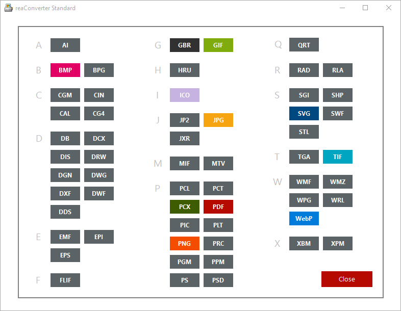
The advent of Optical Genome Mapping (OGM), which utilizes long fluorescently labeled DNA molecules for de novo genome assembly and SV calling, has allowed for increased sensitivity and specificity in SV detection. For example, certain formats can actually contain elements of both types.Whole genome sequencing is effective at identification of small variants, but because it is based on short reads, assessment of structural variants (SVs) is limited. This breakdown isn’t perfect. Most image files fit into one of two general categoriesraster files and vector filesand each category has its own specific uses. Let’s dive into the basics of each file type. Welcome to Image File Formats 101.
Svs File Type Software You Will
NanotatoR uses both external (DGV DECIPHER Bionano Genomics BNDB) and internal (user-defined) databases to estimate SV frequency. The ability to perform script actions in applications can be a very powerful productivity tool that gives customers great flexibility in how they apply Microsoft products to solve real-world problems.We developed an R-based package, nanotatoR, which provides comprehensive annotation as a tool for SV classification. Associating the CZI file.Less obvious examples are file types that allow for embedded script operations, such as Microsoft Access files (.mdb) or macros in Microsoft Word files (.doc) or in Microsoft Excel files (.xls). ResultsDuring the installation of the ZEN software you will be asked if you want to associate the CZI file extension with.
Most quality-control filtration parameters are customizable by the user. From an RNA-Seq dataset, allowing the user to assess the effects of SVs on the transcriptome). If available, expression of overlapping or nearby genes of interest is extracted (e.g. A primary gene list is extracted from public databases based on the patient’s phenotype and used to filter genes overlapping SVs, providing the analyst with an easy way to prioritize variants. Overlap percentages and distances for nearest genes are calculated and can be used for filtration.
We simply add the words INTO OUTFILE, followed by a filename, to the end of the SELECT statement. There’s a built-in MySQL output to file feature as part of the SELECT statement. This may look like an ordinary image, but SVS images are huge: the files are.Save MySQL Results to a File.
SRS platforms used for whole exome (WES) or genome (WGS) DNA sequencing produce billion of reads per run, typically limited in length to 100–150 base pairs (bp). ConclusionsThe extensive annotation enables users to rapidly identify potential pathogenic SVs, a critical step toward use of OGM in the clinical setting.With the advent of the high-throughput short-read sequencing (SRS) techniques, identification of molecular underpinnings of genetic disorders has become faster, more accurate and cost-effective. NanotatoR was also able to accurately filter the known pathogenic variants in a cohort of patients with Duchenne Muscular Dystrophy for which we had previously demonstrated the diagnostic ability of OGM. We evaluated nanotatoR’s annotation capabilities using publicly available reference datasets: the singleton sample NA12878, mapped with two types of enzyme labeling, and the NA24143 trio. NanotatoR passed all quality and run time criteria of Bioconductor, where it was accepted in the April 2019 release. Svg designs bundle, svg design bundle svg shirt bundle quote svg LeeKxstudio 4.5 out of 5 stars (936) Sale Price 3.75 3.75 12.50 Original Price 12.50' (70 off.

Long-read sequencing (LRS) technologies such as nanopore-based sequencing (Oxford Nanopore Technologies) or single-molecule real-time sequencing (Pacific Biosciences) have the potential to both detect complex genomic rearrangements and increase SV break point resolution. Breakpoint resolution is limited by the density of probes on the array.Novel approaches which analyze single, long DNA molecules hold the promise of detecting the previously inaccessible SVs. CMA clinical application is typically limited to CNVs above 25–50 kb, although higher resolution CNV maps have been built and are being used to design disease-specific paths to diagnostic detection of smaller variants (e.g. Chromosomal microarray (CMA) is the established method for high-accuracy detection of CNVs, but it can only identify gains or losses of genetic material and is virtually blind towards identification of balanced rearrangements such as inversions or translocations. Insert size) all affect specificity, sensitivity and/or processing speed of the various variant-calling algorithms. High-complexity regions), noise of data (platform-specific sequencing or assembly errors), complexity of the SV, and library properties (e.g.
The labeled DNA is imaged through nanochannel arrays for de novo genome assembly. For OGM, purified high-molecular-weight DNA is fluorescently labeled at specific sequence motifs throughout the genome (reviewed in ). For example, a comparison of OGM, PacBio LRS and Illumina-based SRS on the same genome showed that about a third of deletions and three quarters of insertions above 10 kb were detected only by OGM. In parallel, a method not based on sequencing, optical genome mapping (OGM, Bionano Genomics), provides much higher sensitivity and specificity for identification of large SVs, including balanced events, compared to karyotype, CMA, LRS and SRS. However, as LRS still isn’t in wide use, SV detection pipelines have seen slower development than SRS-based algorithms, and both quantity and quality of identified SVs vary significantly between tools.
It offers multiple filtration options based on quality parameters thresholds. It determines population variant frequency using publicly available databases, as well as user-created internal databases. Here, we report the development of an annotation tool in R language, nanotatoR, that provides extensive annotation for SVs identified by OGM. Although, OGM is effective in identifying clinically relevant SVs, the currently available SV annotation tools do not provide sufficient variant information for determination of variant pathogenicity. Importantly OGM has allowed refinement of intractable, low-complexity regions of the genome and discovery of genomic content missing in the reference genome assembly. OGM has been effective in identifying pathogenic variants in patients with cancer , Duchenne muscular dystrophy , and facioscapulohumeral muscular dystrophy.
Subsequently, samples were annotated with nanotatoR to examine the performance.Additionally, we tested nanotatoR’s ability to accurately annotate the known disease variants in a previously published cohort of 11 Duchenne Muscular Dystrophy samples. OGM-based genome assembly and variant calling and annotation were performed using Solve version 3.5 (Bionano Genomics). All sample datasets, including “Utah woman” (Genome in a Bottle Consortium sample NA12878), “Ashkenazi family” (NA24143 : Mother, NA24149 : Father, and NA24385 : Son), GM11428 (6-year-old female with duplicated chromosome), GM09888 (8-year-old female with trichorhinophalangeal syndrome), GM08331 (4-year-old with chromosome deletion) and GM06226 (6-year-old male with chromosome 1–16 translocation and associated 16p CNV), were obtained from the Bionano Genomics public datasets ( ). The final output is provided in an Excel worksheet, with segregated SV types and inheritance patterns, facilitating filtration and identification of pathogenic variants.Optically mapped genomes for 8 different reference human samples were used to construct the internal cohort database for evaluating nanotatoR’s performance. It offers an option for incorporating RNA-Seq read counts, which has been shown to enhance variant classification , as well as user-specified disease-specific gene lists extracted from NCBI databases.


 0 kommentar(er)
